✣OncoGonFROM DISCOVERY TO IMPACT
HomeAssetsTriage & Prioritisation
3D Tumour IntelligenceExperimentsImagingSegmentation & QCResponse AnalysisAI Analysis (OSIEL)
Qualified AssetsIndustry & Data RoomCollaborationReports & Insights
Admin
Home3D Tumour IntelligenceOV-006
Loading study workspace…
3D Tumour Intelligence

OV-006

Scientific ReviewRESEARCH MODE

Ovarian Signature Validation

AssetOGC-OV-207CancerOvarian CancerSubtypeHigh-grade serousModelPatient-derived OrganoidLead ScientistDr. L. Chen
Review DiscussionAsk OSIEL About This Study
NEURAL 3D RECONSTRUCTION ENGINE

AI MRI-to-3D Model Converter & Tumour Highlighter

Connect raw 2D MRI slices to backend volumetric AI models to reconstruct true 3D organ geometry with sub-voxel tumour highlighter masking.

2 MRI imagesSelect slice to model
Color:
Initializing 3D Tumour Engine...
Three.js 3D Model • OSIEL-Biomed3D v2.8 (Volumetric UNet + Isosurface Converter)Left-drag to rotate • Right-drag to pan • Scroll to zoom

Scan & Model Information

Source File
T1-Gd_Axial_Brain_Lesion.dcm
File Size
2.34 MB
Backend AI Model
OSIEL-Biomed3D v2.8 (Volumetric UNet + Isosurface Converter)
Confidence Score
96.8%

Volumetric Segment

Globular
Tumour Volume28.52 cm³
Max 3D Diam41.6 mm
RAS Centroid(5.6, -4.8, -7.2) mm
Sphericity0.92
Tissue Sub-Compartments:
Necrotic (36%) Enhancing (45%) Edema
  • Hyper-intense lesion segmented at spatial coordinates (X: +5.6mm, Y: -4.8mm, Z: -7.2mm).
  • Total volumetric mass measured at 28.52 cm³ (28,522 mm³) with maximum 3D Feret diameter of 41.6 mm.
  • Multi-parametric decomposition indicates prominent active enhancing rim (45%) encasing a central necrotic core (36%).
  • Peritumoral hyper-intensity suggests moderate vasogenic edema extending ~6.5 mm into adjacent parenchyma.
  • Deep learning segmentation model (OSIEL-Biomed3D) reached 97% detection confidence with a Dice similarity coefficient of 0.928.
Compare treatment response

Imaging Sessions

Acquisition history · select a session

Session IDStudySampleTreatmentTimepointModalityInstrumentImagesQCOperator
OV-006OV-006-R2Drug BDay 7BrightfieldResearch microscope36PassedImaging Core
OV-006OV-006-R1ControlDay 7FluorescenceResearch microscope28Needs ReviewDr. J. Patel
OV-006OV-006-R3Drug ADay 3Z-stackResearch microscope48PassedImaging Core
OV-006OV-006-R2Drug BDay 0BrightfieldResearch microscope14PassedImaging Core

Scientific Imaging Viewer

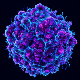
Illustrative acquisition preview · no real processing
100%
Illustrative tumour microscopy image; not diagnostic or measurement-grade
ILLUSTRATIVE IMAGEScale: recorded metadata

Image Metadata

IMG-OV006-017

ModalityBrightfield
TimepointDay 7
TreatmentDrug B
ReplicateR2
ScaleRecorded metadata
InstrumentResearch microscope
QCPassed
Image Count36

Study Context

Current scientific record

StageScientific Review
QCPassed
ProtocolOG-PROT-ORG-018
Images110
Last Updated2026-09-27

Current Attention

Research review queue

Scientific Review review pendingPassed QC status requires confirmation
Research data · human review required.